 Last month, I presented the case of a patient that experienced diurnal variation in vision post-RK. He experienced clear vision in the morning, which got increasingly blurry as the day progressed. Fitting him with a scleral GP lens provided excellent vision, comfort and visual stability. In this month’s column, I want to introduce his daughter who is also a post-RK patient, but that is where their common ground ends. However, despite the differences in their symptoms and outcomes, a similar approach worked for both father and daughter.
A Case Study

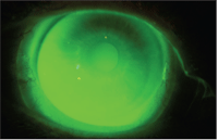
1. RK incisional scars extending into visual axis O.D.
T.S. is a 56-year-old white female with a history of RK O.U. in 1989. Like her father, she too had visual fluctuations, but as opposed to her father, T.S. woke up to blurry vision in the morning, which cleared later in the day. She had recently tried using soft contact lenses with no real success, and had tried corneal GP lenses prior to that with poor adaption.
At the 9:00 a.m. examination, her vision was 20/80 in the right eye and 20/40-2 in the left eye. Pupils were equal, round, reactive and 5mm in normal room illumination. The anterior segment of each eye appeared normal and healthy, with eight radial cut scars in each eye—all of which breached the pupil in normal illumination (figure 1). Her refraction was measured to be +1.25 +.75 x 173, correcting her to 20/30 in the right eye, and +1.00 +1.00 x 5 correcting her to 20/25 in the left eye. Orbscans were also done, demonstrating a typical post-surgical corneal curvature in each eye (figures 2 and 3). Intraocular pressures were measured at 14 in each eye by tonopen at 9:15 a.m.
Because of the great success her father had with scleral lenses, we quickly moved past the discussion of options and initiated a scleral lens fitting for each eye. Our initial strategy of fitting her with an 18.0mm lens design, however, was challenged by symptoms of poor comfort. Despite attempts at changing lens parameters and designs, a larger diameter lens was too uncomfortable.
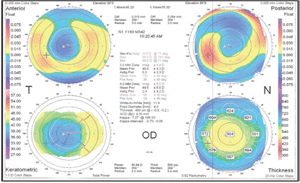
2. Orbscan of the right eye.
Next, we decided to try a smaller diameter scleral design. Based upon the fitting guide recommendations, we placed a 4.40 Sag, 15.8 MSD design on each eye. The fit with this lens demonstrated slight edge lift and awareness with light central touch. We then moved to the 4.60 Sag lens on each eye, which appeared to be a better fit, though there was still a hint of insufficient apical clearance superiorly (figure 4). To help make a better decision on the proper lens, we tried a 4.80 Sag lens on each eye as well, which appeared to be deeper than needed and began to exhibit signs of sectoral compression. Over-refracting the 4.80 Sag lenses produced a final RX of -6.00 and -6.50 in each eye, though best-corrected acuity was only 20/30+ in the right eye and 20/25 in the left eye.
We stopped at this point and had a discussion about whether scleral lenses could achieve the correction she was looking for, as her best-corrected acuity with the lenses was not a significant improvement over her spectacle correction. Often at this point with patients who have irregular corneas, I will darken the room lights and have the patient look at a white target against a black screen to simulate a glare situation. Under these conditions, compared to her spectacle correction, she did notice a significant improvement. Combining these findings with the idea of stable vision throughout the day (which glasses could not provide), T.S. wanted to move ahead with the lenses. We ordered her a pair of lenses with the following parameters:
• R: MSD /4.70 Sag/7.40 profile/-6.00/15.8/non-fenestrated
• L: MSD /4.70 Sag/7.40 profile/-6.50/15.8/non-fenestrated
The lenses were dispensed, and the vision and fit was as expected. Instruction on lens insertion, removal, wear and handling went well, and the patient left wearing the lenses. She has subsequently returned for follow up happy with the lenses and using them several hours a day each day.
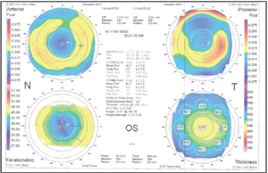
3. Orbscan of the left eye.
Discussion
This case differs from last month’s in several aspects. First, T.S.’s father had clear vision in the morning and became myopic as the day progressed. In this case, T.S. began the day hyperopic, and the hyperopia lessened over the course of the day. In each instance, the eyes moved toward myopia within a matter of hours after waking indicating that the cornea was steepening, most likely due to rising intraocular pressure within hours of waking.
These findings correlate with the PERK study results, which demonstrated that IOP increases as the day progresses, contributing to corneal topographical fluctuations, which lead to an increase in myopia.1,2 By utilizing a scleral lens, which vaults the cornea, the corneal topographic changes are neutralized.
Second, T.S. did not get 20/20 vision through her scleral lenses. This is probably attributable to her larger pupils, which let in more light through the RK incisions and created more blur. It might also be a factor of her RK incisions extending farther into the visual axis. Regardless, the improvement in visual phenomena, such as glare and ghosting, was still substantial enough to make her thrilled with the improvement.
4. Lens fit of 4.60 sag lens on the right eye.
Third, the comfort level with the lens diameter was different between the two. The reason T.S. was having a difficult time with the comfort of the larger lens diameter may partially be attributed to some low-lying conjunctival changes nasally in each eye. By moving to a smaller diameter, we were able to have the lenses put less pressure on these conjunctival areas while keeping the lenses large enough to still maintain corneal vault.
1.Kemp JR, Martinez CE, Klyce SD, et al. Diurnal fluctuations in corneal topography 10 years after radial keratotomy in the Prospective Evaluation of Radial Keratotomy Study. J Cataract Refract Surg.1999 Jul;25(7):904-10.
2.McDonnell PJ, Nizam A, Lynn MJ, et al. Morning-to-evening change in refraction, corneal curvature, and visual acuity 11 years after radial keratotomy in the prospective evaluation of radial keratotomy study. Ophthalmology. 1996 Feb;103(2):233-9.

