Keratoconus is best described as a family of ectatic corneal dystrophies characterized by loss of collagen, corneal thinning and a weakening of structural integrity. With aggressive progression, these changes result in the alteration of corneal morphology that can lead to the loss of best-corrected acuity and eventual visual disability.
| |
| Fig 1. Asymmetric bow-tie pattern with skewed radial axis. | |
 | |
| Fig 2. Pentacam tomography showing early keratoconus. |
Traditionally, practitioners managing visually significant keratoconus have been somewhat limited in therapeutic options. Primary visual rehabilitation methods consist of progressive contact lens options and modifications such as custom toric GP lenses, hybrids, “piggy-back” fits and scleral lenses. However, with potential contact lens intolerance and recurring keratitis due to surface allergy and dry eye, patients can develop anterior corneal scarring and diminished acuity, prompting surgical referral for keratoplasty.
Now, recent developments in surgical management options offer fresh hope for the more severely-affected keratoconus population, while new diagnostic technologies can now uncover the earliest of corneal ectasia alterations. Such cutting-edge detection can allow doctors to refer young patients for collagen crosslinking therapy (CXL) that can arrest the ectatic process, thus saving those affected from a lifetime of visual disability. We have reached a point where the real possibility of early diagnosis and CXL management can offer the hope of halting the advance of this disease.
Early Diagnosis is Key
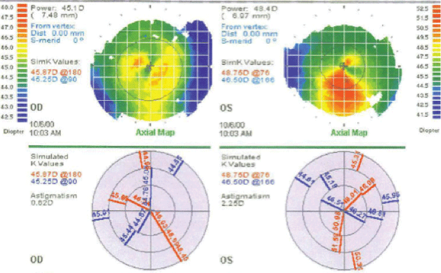
Patients with early changes suggestive of keratoconus often present for routine evaluation. Irregular retinoscopy reflex and red reflex, progressive “against-the-rule” astigmatism and loss of best spectacle-corrected visual acuity are all red flags that should alert one to the possibility of nascent corneal ectasia. Corneal topography revealing inferior corneal steepening or irregular non-orthogonal bow-tie astigmatism patterns and thin corneal pachymetry readings of 500µm or less is also essential in the diagnostic process as well (Figure 1).
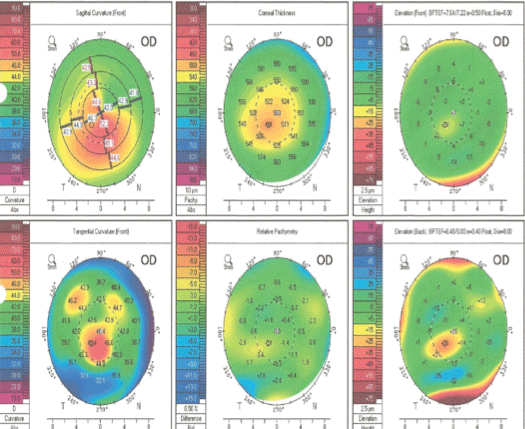
It is crucial that such patients be referred to a center where testing with elevation tomography (Figure 2) can be performed, as it can often reveal subtle elevation changes on the posterior cornea that might not otherwise be apparent. Such changes can be an early harbinger of the ectatic process; clinical suspicion in the examination room is key.
Collagen Crosslinking
The seminal work of Theo Seiler demonstrated that when the cornea was exposed to a narrow spectrum of UVA light after saturation with 0.1% riboflavin (vitamin B2), a process of photoactivation could produce a chemical reaction that increased covalent crosslinking bonds between tri-helical collagen strands and lamellae.1 This crosslinking phenomenon reportedly resulted in a 300% increase in corneal stiffness.
|
|
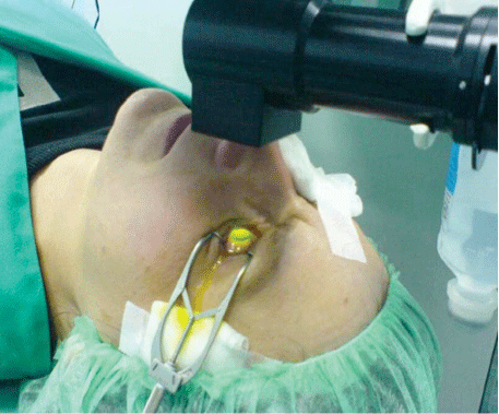
| Fig 3. A patient undergoing epi-off collagen crosslinking. |
Long-term data on patients so treated in the early investigation of CXL showed that 81% to 100% displayed no further progress in the ectasia process.2-4 At our clinic, a three-year experience of treating patients using the epithelium-off approach has thus far documented a complete halt in progression in young patients presenting in the early stages of aggressive ectasia.
Our patients are treated in our refractive surgery suite, where 7mm of central corneal epithelium is removed after exposure to diluted alcohol. Riboflavin 0.1% drops are then instilled every two minutes for 15 applications. Prior to UVA treatment, corneal thickness is checked for a minimum of 400µm to assure endothelial safety. A bandage contact lens is placed on the eye for five days following treatment, and drops consisting of prednisolone acetate 1% and a fourth-generation fluoroquinolone are advised.
Currently, a debate exists as to whether “epi-on” CXL is as effective as the classic “epi-off” approach that we use in our clinic. Our feeling is that complete saturation of corneal stroma with riboflavin and the exposure of the cornea to oxygen is extremely essential to a safe crosslinking process.
Post-op management is similar to that of PRK patients. The treatments have been well tolerated and uncomplicated; however, pain, photophobia, poor healing response and infection have been reported as possible side effects.4
It is important to note that while crosslinking has been reported to improve BCVA by creating corneal flattening and reducing irregular astigmatism, the treatment is primarily aimed at halting disease progression, and patients should be so educated.5 Those with advanced disease are often poor candidates for crosslinking alone; once the disease has fully expressed itself, other options must be considered in conjunction with CXL for ideal results. Currently, CXL awaits final FDA approval in the US.
| |
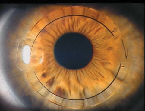
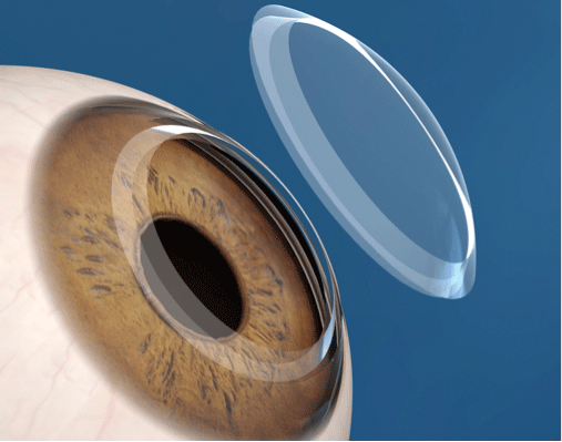
| Fig 4. Intacs rings placed in a keratoconic patient. |
Intrastromal Rings
The Intacs technology was the brainchild of Oklahoma optometrist Gene Reynolds, who developed it as a treatment for low myopia in 1978. Placement of these semi-circular polymethylmethacrylate rings of varying thickness in the cornea was found to flatten corneal radius of curvature. However, the advent of excimer laser in the treatment of myopic refractive errors displaced the technique until 2000, when French ophthalmic surgeon Joseph Colin recognized that selective ring placement could also have a beneficial effect in regularizing corneal morphology when applied to corneas with ectatic dystrophies.6
In a prospective study by Kartakis and associates in 2003, 33 eyes of 26 keratoconus patients received two 0.45mm segments.7 The average follow-up was 11.3 months (ranging from one month to two years) and the mean uncorrected acuity improved from 20/160 to around 20/50. Two eyes lost a line of uncorrected vision, three stayed the same and 28 experienced gains of one to 10 lines. The mean BCVA improved from around 20/40 to a little better than 20/32. Four eyes lost one to two lines of vision, while 25 eyes gained one to six lines. Intacs surgery for keratoconus was approved by the FDA in 2004.
Modern surgical approaches in the use of Intacs rings have now evolved to the point where they have become a viable option in our approach to rehabilitating patients with visually significant keratoconus. When properly performed using femtosecond laser technology, patients with symptomatic keratoconus can obtain better uncorrected “walking around” vision, improved spectacle-corrected visual acuity and better contact lens tolerance.
|
Intacs Performance in Practice
Case 1: Asymmetrical Oval Type A 43-year-old male presents with asymmetric keratoconus. A surgical procedure implanted a single ring 0.45mm at axis 65 with simultaneous collagen crosslinking. 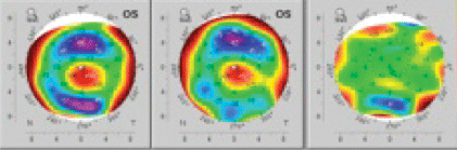
Fig 5. Asymmetric difference map.
Case 2: Symmetric Round Type A 26-year-old female presented with symmetric keratoconus and contact lens intolerance. The surgeon used symmetrical 0.45mm rings with collagen crosslinking. 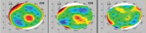
Fig 6. Symmetric difference map.
|
Matching Patients and Procedures
Typically, when I evaluate patients referred for keratoconus, I approach each case from two distinct standpoints: risk of progression and degree of visual impairment. Those early in the disease course are best addressed with CXL alone, but we advise those with significant visual function issues to consider Intacs ring placement in concert with CXL as the two have a synergistic effect in surgical outcomes.
A clinical evaluation of a keratoconus referral consists of a complete examination, including Pentacam tomography to better delineate the pattern of ectasia and corneal thinning. Evaluation of the corneal red reflex with a direct ophthalmoscope allows for the characterization of ectasia patterns as central symmetric (round type) or asymmetric (oval type). Such findings are essential in planning the appropriate custom surgical approach. Symmetrical surgery consisting of insertion of two rings on the steep meridian with round central cases and asymmetric (varying thickness) or single ring placement is typically used in the approach to oval sagging pathology.
In our refractive surgery suite, the Intralase femtosecond laser is used to create ring channels at a depth of 75% of the thinnest corneal pachymetry at a radius of 7mm around the visual axis. The entrance incision is placed at the steepest corneal axis as determined by Pentacam tomography and manual placido ring topography. Following ring placement, CXL is added in the manner outlined above to enhance the flattening effect of ring placement. Patients can expect to experience an evolution of visual improvement over a period of two to three months.
In general, patients with symmetrical findings and corneas with mean keratometry of less than 58 diopters can expect good response when treated with conventional Intacs rings.7,8 Those with corneas steeper than 58D do not respond as well, but remain viable candidates if expectations are limited to achieving better contact lens tolerance.
These patients may benefit from specialty Intacs devices manufactured with an SK modification, which are placed at a 6mm optical zone and have demonstrated a significant improvement in patient response. Unfortunately, they are currently unavailable and awaiting FDA approval.
In our clinic, individuals with sagging oval asymmetric pathology have been shown to do well with single or asymmetric ring placement with mean keratometry readings of up to 60 diopters. Individuals with significant scarring are not good candidates for Intacs surgery except in rare cases where, in conjunction with CXL, goals limited to corneal stabilization and contact lens modification are anticipated. For those patients outside of Intacs treatment range, consider femtosecond laser-assisted keratoplasty.
Femtosecond Laser-Assisted Keratoplasty
Perhaps the greatest advance in corneal transplant surgery has been the addition of femtosecond laser technology. It completely altered the landscape of laser vision correction by allowing surgeons to create predictable LASIK flaps that could be customized to the architecture of each eye. The resulting planar flaps, when used in combination with custom excimer laser ablations, has allowed for superior safety, stability and visual outcomes.
Prior to the advent of laser-assisted keratoplasty, patients having transplant surgery with standard mechanical trephine or mechanical punch transplant surgery could expect a lengthy period of visual rehabilitation upwards of one year. Outcomes notoriously included high levels of irregular astigmatism and wound instability. With femto technology, however, these concerns are no longer viable and the once-dreaded keratoplasty procedure has improved in both recovery speed and outcome predictability.
| |
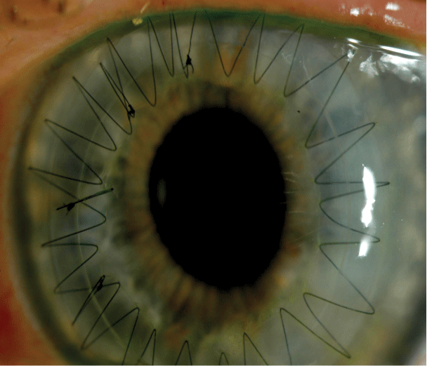 | |
| Figs. 7 & 8. Zig-zag pattern intralase-enabled keratoplasty. |
In femtosecond laser keratoplasty, the corneal surgeon can either calculate a unique pattern of corneal cuts or work off a set group of pre-calculated templates that best fit the patient. Participating eye banks will make use of the same corneal cut parameters to prepare donor tissue. In the operating room, recipient pathology is removed and the custom corneal replacement sutured in place. The precise match of donor and recipient allows for a number of very significant improvements in patient rehabilitation and outcomes. Furthermore, we have found that when a zigzag wound profile is used, increased wound healing interface creates a distinct improvement in wound stability (Figures 7 and 8).
Currently, a series of 14 consecutive keratoconus cases performed over a one-year period with the Intralase femto laser zigzag patterning is under study in our clinic. We have thus far documented visual rehabilitation during a three- to six-month follow-up period. The series consisted of patients ranging in age from 22 to 62 with BSCVA range of CF to 20/100. Three-month post-op data revealed 100% of cases with spectacle-corrected visual acuity of 20/40 or better, with 64% achieving correctable vision of 20/25 or better. In Intralase-enabled keratoplasty (IEK), precise matching of complex donor and recipient tissue planes has allowed for accelerated healing, enabling the removal of sutures as early as six weeks post-op with more than 75% of patients manifesting very acceptable levels of regular astigmatism (2.5D or less.)9
Femto laser technology can also allow for safe astigmatism “touch-ups” for patients with higher levels of postoperative astigmatism. Using the anterior side-cut setting of the Intralase, we can place very safe, accurate astigmatic keratotomy arcuate incisions inside of the graft/host interface to address any astigmatism concerns.10
Summary
Surgical management of keratoconus has clearly reached a point in time where we can offer new hope to this population of patients. To best share these developments, it is incumbent upon all providers of primary eye care to be well acquainted with the options now available.
Of greatest importance is early diagnosis, as patients who present at early stages of the ectasia process require referral and intervention. Primary eye care providers need to be aware of the warning signs of early or progressive keratoconus and be prepared to participate in comanagement.
Timely CXL has been demonstrated itself to provide the opportunity to halt disease progression in young patients who might otherwise be passed over. For those KCN patients unable to tolerate contact lens correction, precise placement of Intacs intrastromal rings can reshape corneal morphology, allowing for improvement in BCVA as well as contact lens tolerance. Patients presenting with advanced disease should be considered for laser-assisted keratoplasty, which offers the prospect of rapid recovery of quality vision and corneal stability.
Dr. Fox is the Medical Director of the Corneal and Refractive Surgery Practice of New York and Clarity Refractive Services of West Orange, NJ. He has over 30 years of experience in refractive surgery as a surgeon and investigator. He can be reached at 917-207-3147 or by email at
[email protected].
1. Spoerl E, Huhle M, Seiler T. Introduction of cross-links in cornel tissue, Exp Eye Res. 1998;66:97-103
2. Caporossi A, Mazzotta C, Baiocchi S, Caporossi T. Long-term results of riboflavin ultraviolet A corneal collagen cross-linking for keratoconus in Italy: The Seina eye cross study. Am J Ophthalmol. 2010;149:585–93.
3. Raiskup-Wolf F, Hoyer A, Spoerl E, Pillunat LE. Collagen crosslinking with riboflavin and ultraviolet-A light in keratoconus: long-term results. J Cataract Refract Surg 2008; 34: 796–801.
4. Ophthalmic and Physiologic Optics, Volume 33, Issue #2, pp78-93, March 2013
5. Kent C. Review of Ophthalmology, Vol. 14, No. 35, September 2014
6. Colin J, Cochener B, Savary G, et al. Correcting keratoconus with intracorneal rings. J Cataract Refract Surg. 2000;26:1117-1122.
7. Bethke WC. Corralling Keratoconus with Intacs. Review of Ophthalmology, Vol. 14, No. 35.
8. Fox ML. Femtolaser Intacs in the Management of Keratoconus: A Stepwise Approach to Successful Outcomes. Ophthalmology Times, publication pending.
9. Fox ML. Laser Assisted Keratoplasty–the Changing Paradigm. Eye World News, Publication pending.
10. Fox ML. Femtosecond astigmatic keratotomy before LASIK can be beneficial. Ocular Surgery News, US Edition, August 25, 2014.

