 |
A 68-year-old Caucasian female contact lens wearer presented for an emergency evaluation on a Sunday evening with a complaint of severe pain in one eye. Her history was significant for an eight-day treatment of that eye with topical Vigamox (moxifloxacin, Alcon) QID and an unspecified steroid drop BID. According to the patient, she initially responded well to the aforementioned treatment, but had since developed a case of intense ocular pain and mild vision loss over the past 24 to 48 hours.
A physical examination revealed the right eye to have normal visual acuity at 20/20- with correction. The left eye with correction was 20/50, pinholing to 20/25-. An external exam revealed a notable area of conjunctival inflammation and hyperemia located in the inferior nasal sector. The remainder of the conjunctiva was relatively clear. An anterior chamber reaction was present, but less than 1+, and the globe demonstrated a remarkable tenderness to touch when palpated through the lid, with the patient indicating pain when the lid was retracted for slit lamp assessment. The cornea displayed a well-defined 2.5mm epithelial defect with no associated infiltration or thinning of the tissue. No purulent activity was detected, and defect margins were soft, but well defined. A dilated examination using tropicamide and homatropine showed no evidence of vitreous cells, and the homatropine provided relief of the pain.
The patient was advised of possible etiologies and alternate treatment options, as the lesion had shown no improvement after a week of therapy. Scrapings from the patient were cultured on chocolate, blood, thioglycolate and Sabouraud’s media and plated on slides. These were delivered to the laboratory for assessment of cellular constitution, as well as resistance and sensitivity. Because of the eight days of prior treatment, expectations for growth were relatively limited; however, I asked that the plates be kept by the laboratory for further evaluation of atypical organisms—specifically, Nocardia, atypical Mycobacterium and other fungi that require an extended growth opportunity to demonstrate their presence.
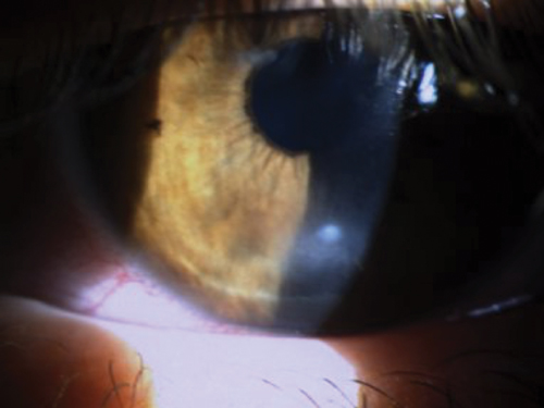 | |
| A minimal ulcer and slight stromal involvement characterized this patient's presentation. |
With regards to the current presentation, potential factors of note included an insufficient dosing regimen, given that current therapy was being administered QID with no significant effect. The possibility that the organism was resistant to Vigamox was also considered; thus, the question was whether to maintain the current agent, add an additional therapeutic item and change the dosage, or switch to a more aggressive therapeutic regimen. Given the late hour of the day and the limited pharmacy options available at that time, I elected to increase the Vigamox to Q2h and add Neosporin ointment TID to the affected eye to increase gram-positive bacterial coverage until fortified agents became available if needed. I also elected to discontinue the steroid that was being administered BID, given the lack of positive response, and add homatropine 5% TID in light of its effect on pain.
The Twist
Two days after the aforementioned change in therapy, no notable improvement in the course or symptoms was observed, though the margins of the lesion appeared slightly softer than previously noted. A trace infiltrate was also apparent below the epithelial defect with no thinning observed. Additionally, the anterior chamber showed trace cell. In light of the relative status quo of the presentation, I elected to continue the recently instituted therapeutic course, but watch the patient carefully. The patient was scheduled for another follow-up exam two days hence, but contacted the office late the following day to advise that the eye had become markedly worse in the last 12 hours, with an increase in redness and discomfort, and a decrease in vision clarity.
Clinical assessment that evening revealed a lesion with minimal increase in the stromal infiltration in the anterior stromal bed immediately below the lesion. The margin of the lesion appeared improved and well-circumscribed, and the tissue was not soft or pliable as before; however, the anterior chamber showed an increase to 1-2+ reaction, with no hypopyon. No significant stromal haze was present outside the area of defect. The globe itself was 3-4+ hyperemic, with an intense zone of ciliary flush.
At that point, I advised the patient that because of the duration of treatment without visible success, I was becoming concerned that this was not a typical bacterial infection, and that the lack of resolution warranted a change in therapeutic intention. I placed the patient on vancomycin Q1hr alternating with Vigamox Q1hr, and discontinued the Neosporin. The patient was atropinized in the office, and then subsequently scripted for atropine on a once-daily basis, given the significant decrease in her discomfort level as a result of the relaxation of the ciliary spasm.
The laboratory was contacted that evening for culture evaluation; no growth was reported. The patient was seen 36 hours after initiation of the additional vancomycin therapy and reported that the eye felt mildly improved. Clinical assessment showed no change in the lesion, although there was a notable decrease in the inflammatory response of the anterior chamber secondary to the use of the atropine.
In the case of lesions of this duration without notable response, the possibility of a viral or other non-bacterial etiology must be considered, though given the appearance of this lesion and the lack of relative neurotrophia on examination, this was unlikely. That being said, the patient was placed on 500mg valcyclovir TID PO and asked to return in two days. At subsequent visits over the following week, the patient showed no notable change. The vancomycin and Vigamox were reduced to QID as a result of the lack of response, and the patient was maintained on oral antiviral therapy. The atropine was decreased to once every other day.
A New Hope
At this point, discussions with respect to the lack of efficacy of any of the interventions, as well as the lack of progression of the lesion and the absence of a positive laboratory outcome from the cultures, led to the decision for a second opinion. Due to the lack of response to a traditional therapeutic regimen, even though the lesion was without satellite involvement and minimal to any infiltrative change or necrosis, the next step was to institute a topical antifungal agent.
The patient was placed on Q2h natamycin therapy with maintenance of topical Vigamox QID and the oral antiviral, and discontinuation of the vancomycin. The laboratory again reported no growth; ironically, however, the patient showed improvement in the first 24 hours on the natamycin. By the third day, the lesion had improved dramatically. It was at this time that laboratory results indicated a Fusarium infection.
The natamycin was maintained with an antibiotic to provide prophylaxis against infection from an opportunistic agent while the eye was compromised by the fungus. The patient’s acuity ultimately returned to almost 20/20 without correction. The anterior chamber was quiet at the last visit, and the lesion was completely resolved.
One interesting aspect of this case: this patient was an avid gardener and, despite the late phase of the season in the north, fungus is not typical; however, the decision to discontinue the steroid and obtain a true picture of the disease state was the key point. In this case, the net change in status was mild, unlike other fungal cases I have managed where discontinuation resulted in rapid progression of the disease state. Having treated one of the first patients in the United States during the Fusarium outbreak several years ago, I requested that the patient bring her contact lenses, lens case and solution to the office to forward to the laboratory for assessment. As of four weeks later, no growth had been identified.

