 |
With the fall season upon us, the potential for adenoviral keratoconjunctivitis (EKC) to present in the office increases dramatically. While the etiology of the disease and its seasonal impact has long been known, recently there has been a significant increase in both the prevalence and severity of epidemic keratoconjunctivitis, one of the forms of the disease, making its management significantly more challenging.
Detect and Distinguish
Singularly, the most important aspect of any treatment is to recognize the presence of the disease and use that knowledge to guide appropriate therapy. A new point-of-care system called AdenoPlus (Rapid Pathogen Screening) permits clinicians to accurately diagnose infectious EKC and differentiate it from many alternative etiologies that can have similar presentations. The system is easily deployable in the office and yields extremely high levels of sensitivity and specificity to the presence of the virus in a patient’s tears.
Capable of detecting all serotypes of the viruses most commonly found in North America, AdenoPlus is particularly sensitive to serotypes 8 and 19.
  |
Typically, a presentation of EKC is relatively mild, with patients showing classic symptoms of hyperemia, tearing, foreign body sensation, subepithelial corneal infiltrates and minor lid edema. Historically, these symptoms are treated with standard regimens, including topical steroids, betadine rinse, ganciclovir, cool compresses and tear supplements. In the last two years, however, a more complex type of infectious disease has presented, with a morbidity that has been profound in some patients. Management of this variant—complex EKC—has proved more challenging due to both its intensity and duration.
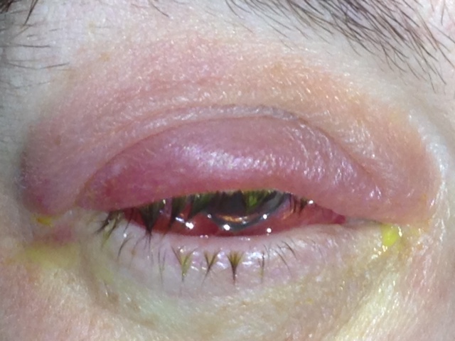
The complex EKC patient presents with significantly greater intensity of disease, including marked periorbital involvement that can include notable spontaneous hemorrhaging, as well as pseudomembranous formation. Additionally, the nummular keratitis typically seen with EKC is much more dramatic and can frequently reduce acuity to the 20/100 to 20/200 range—markedly greater than the 20/20 or 20/25 levels typically seen with the disease. The lid involvement can also be associated with bacterial superinfection and preseptal cellulitis.
Intervene Aggressively
Straightforward and simple to use, betadine has become a relative standard of care for many clinicians dealing with milder cases of EKC. I personally use non-alcohol-based betadine, which can be obtained in either swab or drop application modalities.
A typical procedure is as follows: first, I instill several drops of topical anesthetic and apply the betadine to both the upper and lower cul-de-sacs. I then wait several minutes while the betadine takes effect before rinsing it from the ocular surface with artificial tears and instilling several drops of topical steroid to decrease the somewhat inevitable foreign body sensation that the patient presents with following application.
The use of ganciclovir 0.15% (Zirgan, Bausch + Lomb) is also relatively common, although the clinical science surrounding it is somewhat anecdotal in nature. The first study presented a cohort of just 18 subjects, but demonstrated significant clinical improvement compared to nonintervention, which stimulated the current increasing use of this protocol. Zirgan is used five times per day, and is most effective when initiated within three to four days after the onset of clinical symptomatology. In the more complex cases presented to the office last year, however, Zirgan seemed notably ineffective in managing ocular symptomatology or diminishing the duration or intensity of the disease.
With this recent shift in EKC severity, several traditional diagnostic elements have changed dramatically. First is the bilaterality of the presentation. Traditional thought holds that EKC is a bilateral disease and that other viral entities such as HSK/HZK are unilateral. However, the last two EKC seasons showed over 90% of the patients with RPS-positive findings having 4+ unilateral disease state with no involvement whatsoever of the fellow eye after four to six weeks of follow-up in most patients.
A second significant variation was the rate of bacterial superinfections that presented as preseptal cellulitis. This is differentiated by the level of pain/tenderness on palpation across the lid and the turbidity or tenseness of the tissue. In typical viral disease, the edema is soft and rarely has associated pain on palpation. Of the more than 70 patients who demonstrated severe clinical findings, 18 had concurrent preseptal cellulitis. When managing preseptal cellulitis, I prefer to use a standard penicillin, such as Augmentin 875mg PO BID for 10 days, in combination with hot compress therapy for the first several days to bolster the impact of the antibiotic on the bacterial superinfection. If the patient has a penicillin allergy, either assumed or noted, the use of a first- or second-generation cephalosporin is preferable in light of its excellent Gram-positive coverage.
Another important point when evaluating more severe cases is the need for a thorough examination of the inferior and superior fornices for the presence of pseudomembranous changes. Pseudomembranes often form within the first several days of onset, and will persist and increase if left untreated during the acute phase of the illness. Therapy is directed toward debridement and then subsequent intensive steroid therapy to prevent recrudescence of the lesion. My typical approach is to place several drops of anesthetic in the eye, then soak a cotton swab with phenylephrine and apply it directly to the site. After three to five minutes, a relative hemostasis is achieved that will allow for easier removal of the membrane.
In patients in which the membrane is loosely adherent, a hard cotton swab can sometimes be used to remove the tissue; however, in most cases, a forceps removal is required to tease out the delicate traction that takes place as these lesions develop and cause folding of the conjunctival tissue into layers.
My general preferred technique when there are cicatricial changes present is to use a blunt-tipped element, such as a punctal dilator, to slowly splay the folds open without producing damage to the underlying vascular bed. Following that, I will use forceps to gently tease away the membrane. If the tissue is severely embedded into the conjunctiva, I will try to remove as much as I can without significant damage, then treat the patient aggressively during the next 24 hours with topical steroids to melt down the membrane and allow for easier removal the second day. Membranes on the tarsal plate of the superior lid are sometimes extremely difficult to tease away and, as such, should be managed consecutively over several days.
|
How to Stop An Outbreak
While managing the ocular complications is a critical part of the success in the disease state, the most significant component of good disease management is infection control, both in the community and within the practice. Be mindful of potential transmission by office equipment like tonometer tips and slit-lamp chin rests. Due to the severity of last year’s outbreak, we instituted a universal precaution policy in which any patient who presented with a red eye was isolated until proof with RPS testing was completed. If they tested positive, the room was thoroughly sterilized before further use by another patient. All contact with individuals with red eye presentation was also managed with sterile gloves and disposable sterile equipment to decrease the possibility of transmission within the office. Interestingly, while Zirgan was minimally effective in treating the severe cases, a new protocol in which I prescribed Zirgan for family members of patients with confirmed disease, combined with good home hygiene and infectious disease protocols, was unexpectedly successful: I did not have a single family member of a confirmed infectious patient develop the disease during our treatment periods. |
Following removal, I place the patient on a high potency topical steroid (typically Q1 or Q2 hours) for the next 24 hours, and have the patient return either daily or every other day until the membranes are no longer forming and I have completed their removal. This usually involves one or sometimes two visits; however, some individuals in last year’s group of patients required four to six over the course of eight to 10 days of treatment.
Managing the patient’s nummular keratitis can also be particularly challenging in complex cases. The level of steroid use is intensive and patients have to be monitored for typical side effects associated with that treatment, but abrupt cessation can trigger rebound nummular keratitis. Adding a topical antihypertensive agent can mitigate concerns related to a short-term rise in IOP.
In cases of rebound nummular keratitis, the recurrence can actually exceed the initial treated disease state in some patients. Several individuals in last year’s group of patients developed calcific-like deposits around the virions in the cornea after cessation of steroid therapy, and subsequently had to be restarted on steroid treatment that has persisted in some instances for several months following the initial disease state. Two of these patients had severe enough deposits even following aggressive steroid treatment that PTK is being considered in order to reduce the scar formation and improve visual function.
The management of complex EKC also presents one of the rare opportunities for clinicians to become infected by their patients. This is a significant problem, and in this last year’s epidemic, I treated numerous medical professionals for EKC who been exposed in-office but had not taken appropriate precautions to prevent disease acquisition (see “How to Stop an Outbreak”).
Your role in management—at both the individual patient level and also as an expert resource to the health care community—should be to educate patients on their risk to others, and the need for appropriate hygiene and avoidance of contact with family, friends and coworkers. This is a critical component in minimizing its impact and decreasing the long-term complications of the disease.

