Published February 15, 2017
Retained PK Sutures
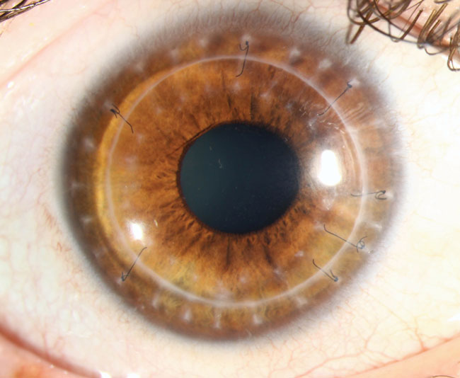
This graft patient showed suture fragments on follow-up. Is it a cause for concern?
Shown here is a 26-year-old female patient who previously underwent penetrating keratoplasty for Acanthamoeba keratitis (AK) unresponsive to medical therapy. The graft size is larger than average due to the advanced state of the AK ulcer and the need for sufficient border zone of healthy, unaffected tissue. She is two years out from the transplant with all her corneal sutures removed, although the broken suture remnants visible here will permanently remain in her cornea. Healing response to graft placement is positive, with no evidence of rejection or failure. The corneal scarring and suture fragments will not affect vision. However, the retained suture fragments could provide a vector for microbial infection, requiring heightened vigilance from both the clinician and patient.
CALL FOR ENTRIES: Do you have great clinical images of fascinating cases you would like to share with your colleagues on this page and online? Send large, high resolution photos (corneal disease or contact lens wear only) and a brief case description to:
[email protected].


