 Scleral lenses are a hot topic. Because scleral lenses are designed to fit the sclera and completely vault the cornea, it takes the irregular corneal surface out of the picture. Even on a patient with keratometry readings over 70D, you’ll have a contact lens that is stable and fits comfortably. But what happens when the sclera is not regular?
Eye care practitioners see scleral and conjunctival anomalies during primary care evaluations everyday. A pinguecula is the most common irregularity encountered during a scleral lens fit. Patients typically report that their eyes get very red in the sector of the pinguecula after several hours of lens wear. The compression of the vasculature under the lens during wear causes congestion of the surrounding blood vessels. The eye stays injected after removing the lens until the congestion of blood has regulated itself.
Case Report
A 52-year-old Hispanic male was diagnosed with pellucid marginal degeneration two years prior and was unable to achieve a satisfactory corneal gas permeable contact lens fit. His GP lens was an intralimbal design, but lacked sufficient movement. He was very comfortable in his soft toric contact lenses, but his vision was unsatisfactory. He was referred for a scleral GP lens fitting.
The scleral lenses provided great comfort and vision, but he complained of chronic redness in the nasal conjunctiva of both eyes. Upon examination, it was evident that the redness was caused by vascular congestion from the scleral lens on his nasal pingueculae.
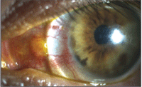
1. Mechanical irritation on a pinguecula induced by a scleral contact lens.
We first attempted some minor changes to relieve the irritation. We ordered the scleral lenses in a smaller overall diameter in an effort to land the scleral lens inside the pinguecula. While this eliminated some of the vascular congestion, the lens was now mechanically irritating the pinguecula (figure 1).
Next, we attempted to vault over the pinguecula with the scleral lens, which relieved some of the irritation, but the lens was difficult for the patient to insert and remove correctly (figure 2).
After failing to resolve the redness, my lab consultant suggested notching around the heaped tissue. A notch in a scleral lens can be effective in eliminating conjunctival irritation from the overlying contact lens by contouring around scleral irregularities.
There are several ways to measure and order a notch in a scleral contact lens. One way to begin is to simply dot the lens with a marker around the scleral irregularity while the patient is wearing the lens. After the lens is removed, measure the height and width of the dotted area.
Another way is to use a parallel-piped light beam in the slit lamp and measure the height and width of the pinguecula under the contact lens.
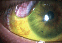
2. Incorrect application of a scleral lens leading to a bubble within the corneal chamber.
For insertion, instruct the patient to apply the lens with the notch in the corresponding quadrant, and it will settle into place. Patients will notice improved comfort and cosmesis, leading to a longer wearing time. Our patient immediately noticed improved comfort and cosmesis (figure 3).
With all of the customizable options available for scleral lenses, patients with specialized vision needs no longer have to settle for a lens that just “gets them through the day.” Notching a lens to improve scleral contour is one way to customize a scleral lens for your already unique patient.
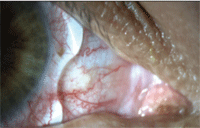
3. A notched scleral contact lens with improved fit and appearance.

