 To keep our patients in their contact lenses, we need to leverage every tool that we have in our practice. Sometimes this means thinking of outside-the-box uses for traditional equipment.
In this month’s column, we explore alternative uses for corneal topographers, and discuss how the results of these tests may give us an enhanced understanding of contact lens dynamics to maximize successful lens wear and minimize dropouts.
Understanding the Line of Sight
Fitting soft multifocal contact lenses present unique challenges. There are times when two patients with very similar ocular characteristics may have very different outcomes with multifocal lenses. Some patients’ visual complaints may be fixed by following the multifocal fitting guide, while this does very little to improve the visual outcome for others. There are times where we dismiss these failures or simply attribute them to a patient being too “picky” or unable to adapt to a multifocal lens. While this may occasionally be true, there are likely other physical measurements that are contributing to the success or failure of lens wear.
Matthew Lampa, O.D., and colleagues recently presented a poster that looked at the difference between the anatomical center of the pupil and a patient’s line of sight.1 They used a topographer to measure the difference between the center of the pupil and the central ring of the videokeratoscopic image as it is centered over the patient’s line of sight (figure 1). They also measured topographies over the surfaces of multifocal contact lenses. All of the contact lenses that they examined on the patients’ eyes were well centered when viewed with a slit lamp, but some patients did show a poor centration of the multifocal lens optics over their line of sight.
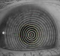
1. The videokeratoscopic image of the right eye shows a prominently displaced nasally located line of sight with respect to the center of the pupil.
Anecdotally, we have been paying particular attention to videokeratoscope images and comparing the difference between the line of sight and pupil center. We’ve also been performing topographies over the surface of aspheric multifocal lenses, and have seen a high rate of success with patients whose lens optics appear to line up well over the patient’s line of sight.
For those patients whose multifocal lens optics are not lined up over their line of sight (even though at the slit lamp it may be well centered), we’ve found it difficult to meet the patient’s visual demands even when troubleshooting with the fitting guides. This is particularly true in higher add powers.
Although additional research to clarify the role of the optics of the lens centering over the patient’s line of sight and its effects on patient outcomes is still needed, this is certainly an important variable that deserves attention and may help better explain successes and failures with simultaneous vision soft multifocal contact lenses.
Topography of GP Surfaces
Just as topographies can be taken over the surface of soft contact lenses, they can also be utilized over the surface of gas-permeable (GP) contacts to better understand the patient’s visual experience.
One such example is a patient wearing a Bi-expert (Baush + Lomb) contact lens, which is a segmented multifocal lens. This lens is unique in that it does not have a visible line on the lens. When the lens is sent to your office, it comes with a small dot that is located on the nasal and temporal aspect of the lens that corresponds to where the line that separates the near and the distance optics are located. This marking usually wipes away within a few weeks through normal care for the contact lens.
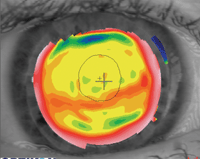
2a. Topography over a segmented bifocal. Note the change in topography profile
corresponding to the segmented portion of the RGP and the pupillary margin with respect to the segment line (left).
When working with a segmented bifocal, also consider these requirements:
• First, make sure that it translates effectively and allows the patient to see through the near optics of the lens when looking down.
• Second, make sure that the segment is high enough to allow easy transition between the distance and near optics, and low enough so that it doesn’t interfere with distance vision. Keep in mind that, while we can get a sense of whether the segment is too high or too low based on changes in vision that the patient describes while tilting their head either forward or backwards, this cannot be visualized at the slit lamp
Topography over the surface of the lens can easily allow visualization of changes on the contact lens surface (figures 2a and 2b). This lets you easily determine the transition zone between the distance and the near optics, and also allows you to observe where the pupil is with respect to the transition zone and whether the segment is low enough in primary gaze. This is valuable for any segmented multifocal GP that does not have a visible line dividing the distance and near optics in the lens.
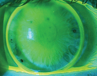
2b. A segmented RGP bifocal lens. The dots at 2:30 and 8:30 represent the edges of the segment of the lens. The dot at 7:30 is to identify the lens as the right lens (right).
Infrared Capabilities
Some topographers offer the unique ability to view the meibomian glands through infrared imaging. These capturing systems can image the meibomian glands of everted upper and lower eyelids. It was recently shown that meibomian gland dropout is positively correlated with the ocular surface disease index score.2 Meibomian gland dropout and tortuosity of the glands, both considered signs of meibomian gland dysfunction, can easily be imaged.
This is an interesting and critical finding especially when working with those who suffer from dry eye symptoms, particularly presbyopes. By viewing the meibomian glands in this way, the practitioner has one more factor to consider when fitting and treating patients for contact lens-related dryness (figure 3).
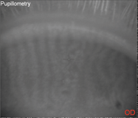
3. A picture of the tarsal plate showing the meibomian glands in a healthy individual.
For example, a patient who complains of comfort issues and has relatively normal ocular health findings, other than visible meibomian gland dropout with infrared imaging, may be best served by optimizing the function of the meibomian glands.
With new technologies come new ways to view contact lenses and the tissues that support comfortable, optimized lens wear. Corneal topographers may provide a unique opportunity to view contact lenses and meibomian glands, which can help to improve patient outcomes and minimize dropouts.
1. Lampa M, et al. Assessing multifocal soft contact lens centration with the aid of corneal topography. Poster presented at the annual Global Specialty Lens Symposium meeting, January 26-29, 2012; Las Vegas.
2. Srinivasan S, Menzies K, Sorbara L, Jones L. Infrared imaging of meibomian gland structure using a novel keratograph. Optom Vis Sci. 2012 May;89(5):788-94.

