 |
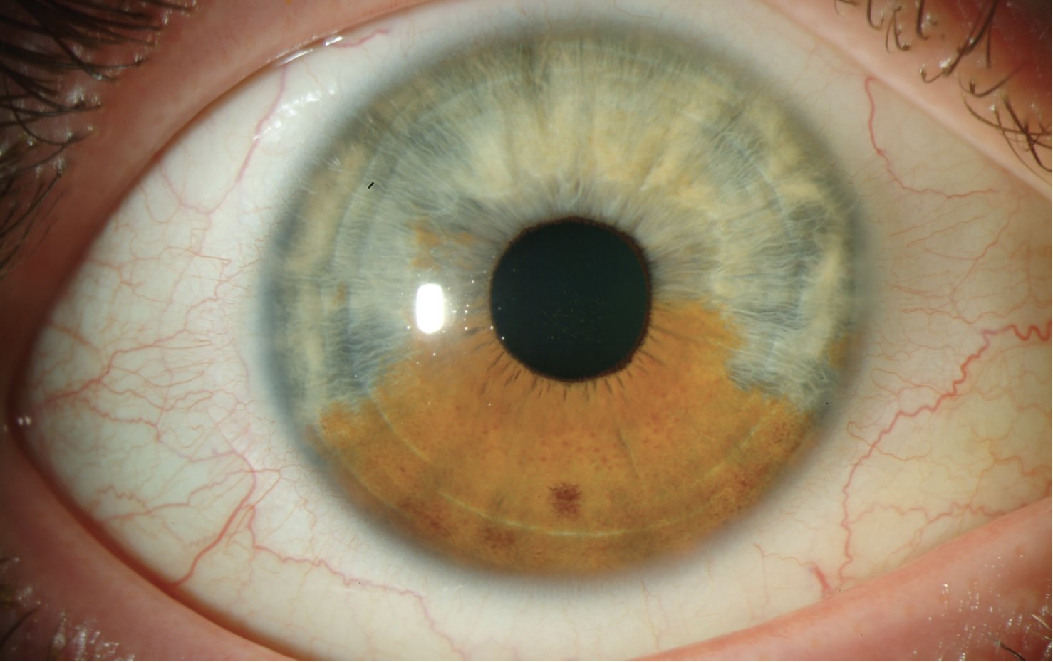
A 26-year-old female presented for a comprehensive exam and contact lens fitting. Her ocular exam was remarkable for a large iris nevus covering the inferior half of her right eye. The patient reported that her eyes have always been two different colors. The pigment appeared shallow with mammillation over the pigmented surface. She did not have a pigmented sclera or pigment around her eyes. She had large optic nerves with 0.8 cupping OD and OS, with inferior and superior nerve fiber layer thinning OD>OS. Her pressures were 17mm Hg OU. Visual fields were full OU.
 |
|
Click image to enlarge. |
The differential diagnosis of iris pigmentation includes iris freckle/nevus, iris melanocytoma, iris melanocytosis and iris melanoma. Iris freckles and nevi are just an increase in melanin pigment and not a precursor to melanoma; however, having three or more freckles is associated with an increased risk of cutaneous melanoma.1 If corectopia or iris ectropion is present, there is an increased concern for uveal melanoma.
An iris melanocytoma will have a large area of brown homogenous pigment with a granular surface, and there may be seed pigment around it. In such cases, there is a small risk for developing melanoma; however, there is an additional risk of developing glaucoma.
Iris melanocytosis is a congenital condition where the uvea gets too much pigment. It can be complete or sectoral, with tiny micronodules (mammillations) in the pigmented area. The sclera and periocular skin may also have pigmentation. There is a 1:400 risk of developing melanoma.1
All iris pigment should be monitored for evidence of growth, vessels in the pigmented lesion, pigment seeding in the angle, evidence of tumor in the ciliary body or elevated IOP. Any pigment covering more than three clock hours is suspicious.
Our patient had a complete glaucoma workup, including gonioscopy, echography of the ciliary body and photodocumentation. She will continue to be monitored at six-month intervals.
1. Laino AM, Berry EG, Jagirdar K, Lee KJ, Duffy DL, Soyer HP, Sturm RA. Iris pigmented lesions as a marker of cutaneous melanoma risk: an Australian case-control study. Br J Dermatol. 2018 May;178(5):1119-27. |

