 |  |
The ocular surface is a remarkable system. When it functions properly, an adequate quantity and quality of tears help the eye remain unnoticed by the person as the lids spread the tears across the surface, allowing light to enter the eye through a smooth refractive surface. And on this normal functioning ocular surface, contact lenses provide a remarkable option for vision correction. But, disrupting this delicate balance can cause increased awareness of the eyes, poor vision, desensitization of the cornea and anatomical alterations such as staining. Even with early stages of ocular surface disease (OSD), a contact lens can potentially compromise the comfort and visual performance the lens was designed to provide.
Although mild OSD can be treated immediately and rarely requires any discontinuation of lens wear, patients often do so on their own due to discomfort. This is where the clinician steps in to treat the ocular surface appropriately and get patients back to their contact lenses.
Dry Eye and CL Wear
Dry eye is at the top of the list of ocular surface disruptions that can wreak havoc on contact lens wear. Luckily, a better understanding of its underlying etiology and newer diagnostic technologies allow us to diagnose the condition early and manage patients’ response to treatment better than ever before.
Clinicians have a number of treatment options to help care for patients with OSD—a must for all patients, but especially to keep contact lens wearers happy in their lenses. Dry eye therapy can be a daunting task, but this two-step approach to optimizing the ocular surface—and thus the lens wearing experience—can help keep you on the path to success.
Step One: Relief
In this two-pronged strategy, clinicians must first provide relief of patient symptoms such as dry, irritated, itching or burning eyes or even blurred vision or fluctuating vision with blinks. Temporarily increasing the volume of tears on the eye with artificial tears is often the first move to alleviate discomfort. The improved function these artificial tears produce will also provide further relief.
For patients requiring significant supplementation of tears, clinicians can consider gels or ointments, always keeping in mind the blur profile these products produce. These typically work well in the evening for patients who have incomplete lid closure, for example. Another option to relieve dry eye symptoms over a longer period is Lacriserts (Bausch + Lomb)—small hydroxypropyl cellulose inserts placed in the lower fornix that slowly dissolve demulcents into the tear film over a 24-hour period.
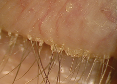 |
| Fig. 1. A patient with prominent anterior blepharitis. |
Step Two: Function
Improving function is just as important—if not more—than initially providing relief. Uncovering and treating the root cause of the condition will ultimately provide more relief to the eye by also improving the function of the ocular surface.
For one, clinicians should assess each dry eye patient’s blink to ensure it is complete. They should also check lid elasticity by examining the lower palpebral conjunctiva and everting the upper eyelid to rule out floppy eyelid syndrome (FES). Individuals with FES may experience symptoms consistent with dry eye from unintended eye opening in the evening and, in some patients, eyelid eversion.1
There are two ways to improve function: mechanical means and chemical means. Only by providing both mechanical and chemical therapies will the functionality of the ocular surface improve. Better function will eradicate dry eye symptoms for many patients, and as rehabilitation progresses, supplemental lubrication can taper. However, some will still require the use of lubrication.
Mechanical. To treat the ocular surface through mechanical means, clinicians must first be confident in the likely cause of the patient’s symptoms. For example, the best treatment for a patient with anterior blepharitis is cleaning the eyelids of excessive debris (Figure 1). For this, clinicians can use the BlephEx (Rysurg) in office to mechanically remove excessive debris at the base of the lashes.
For patients with meibomian gland dysfunction—whether obvious or non-obvious—debridement of the lid margin with a metal spud helps remove keratinized tissue that may be obstructing the meibomian glands (Figure 2). In addition, thermal application can decrease the viscosity of the meibum secreted from the meibomian glands. Patients can use a commercially available warm compress at home on a daily basis, or they can return to the office for an in office thermal therapy such as MiBoFlo (Mibo Medical Group) for the anterior surface of the eyelids or Lipiflow (TearScience) for the posterior surface of the eyelids. Lipiflow also produces pressure through an inflatable air bladder on the anterior portion of the lid to help evacuate meibum from the glands.
For individuals whose MMP-9 assessment by InflammaDry (RPS) testing is unfavorable and are currently being appropriately treated, consider adding punctal plugs. Punctal plugs help retain tears on the surface for longer periods of time by decreasing the rate they drain. However, using artificial tears on eyes with high levels of inflammation is not beneficial. Thus, InflammaDry is a good tool to assess contact lens wearers experiencing dry eye symptoms with low levels of ocular surface inflammation.
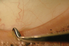 |
| Fig. 2. Lid margin debridement, as seen here, can significantly improve a patient’s ocular surface function. |
Chemical. Clinicians have a number of strategies available to us to chemically improve the health of the ocular surface and adnexa. If, for example, an anterior blepharitis is present and the eyelids have been cleaned in office, having the patient perform lid hygiene on a daily basis with a commercially available preparation is appropriate.
For patients with significant inflammation, a temporary course of topical corticosteroids may be appropriate.
For individuals requiring long-term management of inflammation, Restasis (cyclosporine, Allergan) has been used for well over a decade, as it modifies T-cell interaction through immunomodulation.2 Another recent addition, Xiidra (lifitegrast, Shire), is a topical lymphocyte functioning antigen antagonist that prevents the interaction of T-cells with intercellular adhesion molecules on the ocular surface, ultimately hindering T-cell activation and the release of inflammatory mediators.3
Oral agents can also help improve the ocular surface function. Omega-3 fatty acids, for example, have been long known to have anti-inflammatory activity.4 In the body, this produces increased levels of prostaglandin-3, which has strong anti-inflammatory activity. Additionally, naturally occurring omega-6 fatty acids extracted from evening primrose oil have strong anti-inflammatory activity. Research shows formulas containing both omega-3 and omega-6 fatty acids can be beneficial for patients with dry eye.5
Another oral agent that works remarkably well to help improve ocular surface function through decreasing inflammation is doxycycline. This antibiotic, commonly used in dermatology for treating signs and symptoms of rosacea, has a non-specific anti-inflammatory activity. Thus, patients with ocular rosacea—which causes an inflammation of the meibomian glands and a visible alteration in the meibum secreted—often benefit from long-term doxycycline therapy, as it inhibits MMP-9.
Dry eye is often a remarkably complex condition and, if left untreated, can degrade comfortable contact lens wear. By actively identifying the underlying etiology of dry eye symptoms and appropriately treating it, clinicians can improve patients’ chances of wearing lenses comfortably. A two-pronged approach—provide relief and improve function—is key to strategically caring for these patients.
1. Skorin L Jr, Knutson R. Ophthalmic diseases in patients with obstructive sleep apnea. J Am Osteopath Assoc. 2016;116(8):522-9.
2. Sheppard JD. Dry eye moves beyond palliative therapy. Manag Care. 2003;12(12 Suppl):6-8.
3. Abidi A, Shukla P, Ahmad A. Lifitegrast: A novel drug for treatment of dry eye disease. J Pharmacol Pharmacother. 2016;7(4):194-8.
4. Georgakopoulos CD, Makri OE, Pagoulatos D, et al. Effect of omega-3 fatty acids dietary supplementation on ocular surface and tear film in diabetic patients with dry eye. J Am Coll Nutr. 2017;36(1):38-43.
5. Molina-Leyva I, Molina-Leyva A, Bueno-Cavanillas A. Efficacy of nutritional supplementation with omega-3 and omega-6 fatty acids in dry eye syndrome: a systematic review of randomized clinical trials. Acta Ophthalmol. March 30, 2017. [Epub].