In this era of rapid technologic advances, it can be difficult to keep up with the latest equipment and procedures within the field of eye care. Even when we do stay abreast of these changes, do we need or want to buy the latest instruments? Some practitioners feel that the old standby is just fine, while others are eager to have the newest tools immediately. The path to follow is likely somewhere in between. In this article I look at some of the instrumentation and lenses of the past and present, and discuss the pros and cons of each in relation to contact lens practice.
Visual Acuity
Let’s start with measuring visual acuity. Traditionally, this was done with a Snellen wall chart set 20 feet from the patient. The patient was asked to cover one eye and read the letters, then repeat with the other eye. Of course, there are some pitfalls to keep in mind. First, the lighting must be proper; and second, we have to be conscious that the patient is not already familiar with the letter sequence.
The next step up is the standard projected chart, which ensures good lighting of the letters and good contrast, but we still find that the patient’s memory can be a factor here.
The latest instrument for acuity measurement is a computer-based system like the Smart System II 2020 Wireless Remote (M&S Technologies). With this arrangement, the letter selection and sequence is infinitely varied, thus memorizing the chart is not a concern. Because contrast and lighting can be controlled and varied, contract sensitivity and vision can both be assessed. Personally, having worked with such a system for several years, it is difficult to switch back when doing a vision screening or working in a satellite office without this equipment. Score one for new technology!
Refraction
Most of us are taught to use a phoropter to perform refractive evaluations. We get good at retinoscopy to find a starting point for new patients. In fact, the retinoscope and phoropter have been mainstays for refractive determinations for over 100 years.
But, before the phoropter, there was the trial frame and loose lenses. While the phoropter has the advantage of allowing the measurement of phorias, ductions and AC/A ratios, a trial frame refraction can be just as fast—and sometimes more accurate—than a phoropter refraction. Think about the young, fidgety patient who will not sit properly behind a phoropter, or the older, infirm patient who cannot be aligned behind a phoropter. In these instances, a trial frame refraction is a far better option. Other examples of the superiority of the trial frame include cases when patients present with large ametropias (particularly astigmatism) and when you need to do an over-refraction on bifocal/multifocal contact lens patients. I would give this point to old technology.
When it comes to determining the initial refractive values on a new patient, many practitioners have embraced the autorefractor and moved away from retinoscopy. This allows technicians to determine the objective refraction, and frees up the doctor for other tasks such as subjective refraction. A subjective refraction is still needed after the objective determination to refine the prescription.
Back in the 1970s, Humphrey Instruments introduced the Humphrey Vision Analyzer, which is a fully subjective refracting system.1 It used what was termed a “remote refraction,” involving a system that manipulated lenses before the projector rather than before the patient’s eyes.2 The results were accurate and repeatable—but a cost of $31,500 to $39,000 per unit made it an expensive option for the average practitioner.3
A new refracting system, the PSF Refractor (VMax Vision), uses a point-spread function by measuring a patient’s subjective visual response to a point source.4 This enables correction of higher-order aberrations by giving precise refractive results. Coupling that instrument with VMax’s Encepsion lenses brings the cost of the unit from $25,000 to $45,000, depending on the selected package. Time will tell whether or not the cost proves to be a stumbling block.
Cornea
Examining the cornea requires use of a slit lamp biomicroscope. The basic design has not changed much over the years; there are currently two instrument configurations dominating the market. There is the Zeiss design with a low-profile illumination system and a large magnification range, and there is the Haag-Streit design with a tall illumination tower and a choice of two magnifications (at least in the basic model). Practitioners may debate the merits of one design over the other for clear images, but both work very well, so it really is a matter of personal preference.
In recent years, specular microscopes have been developed to provide a fantastic view of corneal structures. They also offer some advantages in diagnosing infections and other problems with the cornea. While they may be very helpful for evaluating the endothelial guttata before cataract surgery, they are not going to replace the slit lamp for routine evaluations. Also, keep in mind that the cost of the unit can reach $35,000 or more.
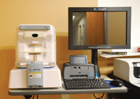
Konan Specular Microscope.
We also need to evaluate the corneal curvature, especially before contact lens fittings. The traditional instrument for this task is the keratometer. These units have not changed much since electricity replaced candles and mirrors for the illumination system. Keratometers measure the curvature of the central 3.5mm of the cornea, which gives us information about that area but not what is going on beyond that.
Corneal topographers, developed in the 1980s, allow for a wider measurement area and give more information than just the curvature of the cornea. They provide a two-dimensional view of the elevation and variation of the cornea to about 12mm. Topographers are great for following keratoconus patients because they allow you to detect subtle changes over time.
A newer instrument for accessing the cornea and other anterior segment structures is the corneal tomographer. The more advanced technology employed in these devices allows a three-dimensional view of both the anterior and posterior cornea, the anterior iris and crystalline lens.5 We also must mention anterior segment optical coherence tomography (OCT), which is even more sophisticated that the previous devices and covers a larger area of the cornea and sclera.6 Its sensitivity allows the practitioner to easily discern the level of the cornea involved in dystrophies, degenerations and infections.7 This is very valuable for the anterior segment/corneal surgeon, as well as fitters of large-diameter gas-permeable lenses.
Contact Lenses
In contact lens practice, there has been tremendous change due to technological advances. Back in 1888, contact lenses were literally glass shells. They had zero oxygen flux and were very involved from a fitting standpoint. They were large lenses that did not move much and caused a lot of corneal edema and conjunctival injection.
The development of polymethyl-methacrylate plastic allowed for smaller lenses to be made, and a more precise lens-to-cornea fitting relationship was achieved. While the material did not allow oxygen to pass through, the smaller size and the ability to move on the cornea did reduce the instances of corneal edema to a more acceptable level. Adding silicone and other components to the polymer mix did increase oxygen permeability and transmissibility. These advances have allowed gas-permeable lenses to be used for extended wear, up to 30 nights, with very few problems. The new material advances also have made it possible to manufacture larger lenses, with diameters above 24mm, enabling better comfort and offering relief from painful ocular conditions. Chalk that up to another point for new technology, and we won’t even talk about the advances in lathing technology that make these new lenses possible. (That would be an article of its’ own.)
Soft Contact Lenses
Soft contact lenses currently make up about 90% of the lenses being used worldwide.8 From their humble beginnings in the kitchen of Professor Otto Wichterle in 1961 to the silicone hydrogel lenses of today, polymer technology for soft lens materials has been steadily changing. The earliest materials, such as polymacon, had very low dK values and did cause corneal edema in some patients. Polymers with increased water content gave some relief from corneal swelling, thus the idea of extended wear was born.
Following an outbreak of corneal infection and ulcers in the mid-1980s, Oliver Schein, M.D., and colleagues conducted the first major study of extended wear complications.9 Clearly embracing new technology can have its own perils. However, the setback spurred new development in polymer technology and eventually resulted in silicone-based polymers that were wearable and had dK values up to 175. These changes allowed for safer extended wear, or continuous wear up to 30 days, but they were not without risks.10 Even daily wear of the silicone-hydrogel materials did not eliminate problems like keratitis, superior epithelial arcuate lesions (SEALs), contact lens-induced acute red eye (CLARE) and infiltrates.11
The changes in technology for manufacturing soft lenses have allowed us to move away from the need to save our lenses for a year to a much more convenient disposable schedule. Early lenses were prone to deposits of denatured proteins on the surface, so the thought was that replacing them more frequently would eliminate that problem and the inflammation issues related to conventional soft lenses. Whether the replacement schedule is every one month, two weeks or daily, complications such as keratitis still exist.12
Lens Care
The evolution of lens care technology has also been tremendous. We went from having to make our own cleaner and saline to selecting from a multitude of laboratory-made products and from heat disinfecting to chemical and peroxide-based systems, all new technologies designed to increase comfort and ease. Yet, we still find issues with irritation and eye redness in some patients—stemming in part from interactions between the polymers and solution components, and in part from patient non-compliance.13
Patient education, and re-education, about the importance of following the proper wear, care and replacement schedules is very important. All things being equal, when it comes to lens materials, using the most oxygen-permeable polymer available makes the most sense. The shortest possible replacement frequency is advisable to decrease the likelihood of complications due to surface changes over time.
The skills and expertise we attain from optometry school to our practice lives and continuing education are enhanced by all the new technology and the studies that support them. We put all of this information to use during our interactions with each patient. Ultimately, we formulate the best plan for the individual in our chair, regardless of whether it involves old or new technology.
1. Efron N. Instrument review: The Humphrey Vision Analyser. Aust J Optom. 1982 Apr; 64(4):149-53.
2. Alvarez LW. Development of variable focus lenses and a new refractor. J Am Optom Assoc. 1978 Jan;49(1):24-9.
3. Kratz LD, Flom MC. The Humphrey Vision Analyser: reliability and validity of refractive-error measurements. Am J Optom Physiol Optics. 1978 Oct; 54(10):653-9.
4. Rosenweig T. Instrument Focus: Provide night sight—refractive device enhances night vision. Optom Management. 2011 Nov;46(11):62-3.
5. Yeung KK, Chang S. New equipment sees the cornea better. Rev Cornea Contact Lens. 2011 Oct;147(7):16-9.
6. Sobara L, Maram J, Fonn D, et al. Metrics of the normal cornea: anterior segment imaging with the Visante OCT. Clin Exp Optom. 2010 May;93(3):150-6.
7. Vajzovic L, Karp C, Haft P, et al. Ultra high-resolution anterior segment optical coherence tomography in the evaluation of anterior corneal dystrophies and degenerations. Ophthalmology. 2011 Jul;118(7):1291-6.
8. Bennett ES. GP Annual Report 2011. Contact Lens Spectrum. 2011 Oct;26(10):28-33,48.
9. Schein OD, Glynn RJ, Poggio EC, et al. The relative risk of ulcerative keratitis among users of daily-wear and extended-wear soft contact lenses. N Eng J Med. 1989 Sep;321(12):773-8.
10. Schein OD, McNally JJ, Katz J, et al. The incidence of microbial keratitis among wearers of a 30-day silicone hydrogel extended-wear contact lens. Ophhalmology. 2005 Dec;112(12):2172-9.
11. Stapleton F, Stretton S, Papas E, et al. Silicone hydrogel contact lenses and the ocular surface. Ocul Surf. 2006 Jan;4(1):24-43.
12. Yeung KK, Forister JFY, Forister EF, et al. Compliance with soft lens replacement schedules and associated contact lens-related ocular complications: The UCLA Contact Lens Study. Optometry. 2010 Nov;81(11):598-607.
13. Carnt N, Jalbert I, Stretton S, et al. Solution toxicity in soft contact lens daily wear is associated with corneal inflammation. Optom Vis Sci. 2007 Apr;84(4):309-15.
Dr. Benoit is senior optometrist with The Eye Center of Concord, a multi-subspecialty ophthalmology group located in Concord, N.H. He is a diplomate and chair of the American Academy of Optometry’s Section on Cornea, Contact Lenses and Refractive Technology.

