 |
A 29-year-old bilateral aphake was referred to the University of Iowa for concerns of corneal edema and decompensation. He had pediatric cataracts removed when he was two months old and has a 29-year history of gas permeable contact lens wear.
On presentation, he showed 360 degrees of microcystic edema with a central clear cornea. He denied changes in vision or discomfort.
He suffers from Brown-McLean syndrome, which is visually astonishing but quite clinically benign. This condition usually presents as peripheral corneal edema in patients with long-term aphakia after intracapsular cataract extraction.1
Intracapsular cataract surgery, an older technique in which both the lens and capsule are removed, is rarely used today. While once the standard of care, today this type of cataract surgery is reserved for cases where the lens has dislocated secondary to injury or disease. Brown-McLean syndrome may also present after extracapsular cataract extraction and phacoemulsification or pars plana lensectomy and vitrectomy.1 Rarely, it occurs in eyes that have not had surgery.1
The corneal edema starts after a latent period of several years and is seen in the peripheral 2mm to 3mm of the cornea, starting inferiorly and extending circumferentially in severe cases. Guttae are present on the endothelium and may have punctate brownish pigment. No neovascularization will be present, and the limbus and conjunctiva are unaffected. The central cornea also remains unaffected, and specular or confocal microscopy will show normal endothelial counts and cell morphology.2
Most patients are asymptomatic, but some may complain of foreign body sensation or even pain from ruptured bullae. Patients are managed with normal contact lens wear or glasses and symptomatic relief of bullae, as necessary, with hyperosmotic agents.
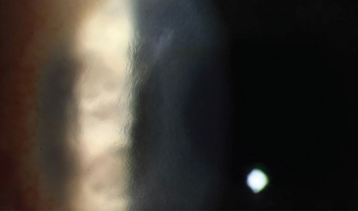 |
| This patient suffers from Brown-McLean syndrome, which is visually astonishing but quite clinically benign. Click photo to enlarge. |
| 1. Gothard TW, Hardten DR, Lane SS, et al. Clinical findings in Brown-McLean syndrome. Am J Ophthalmol. 1993;115(6):729-37. 2. Lim LT, Tarafdar S, Collins CE, et al. Corneal endothelium in Brown-McLean syndrome: in-vivo confocal microscopy finding. Semin Ophthalmol. 2012;2013:6-7. |