 Keratoconus is a condition for which treatment has been limited to palliative intervention during the early phases of the disease, and surgical intervention in the late-presenting cases. This has created a challenge to clinicians and patients alike, in that doctors’ best efforts frequently were just good enough to allow the patient to function visually.
Collagen crosslinking (CXL) brings a new option to keratoconus therapy, and opens the door to a remarkable future for clinicians and patients.
The underlying principles behind crosslinking involve the use of riboflavin in a special mixture that generates increased penetration, along with highly focused ultraviolet light at 370nm. The combination of these elements in the corneal stroma creates a reactive oxide, which structurally enhances the collagen matrix and strengthens the stroma, ceasing the progression of thinning and forward coning of the corneal surface.
The procedure can be applied in a multitude of ways. There has been some controversy over whether epithelial removal (“epi-off” crosslinking) improves riboflavin penetration compared to the “epi-on” technique of leaving it intact. This has been addressed in numerous clinical trials, and is currently under review in the Avedro CXL USA trials. Data shows both interventions to be efficacious, with the difference in outcomes based primarily on the management of the epithelial wound healing in epi-off patients and the relatively small, but real, risk of complications related to wound healing delay and the potential for infectious disease.
CXL is best applied to the keratoconic patient in the earlier phases of the disease state. Typically, the corneal thickness should be greater than 400 microns. Additionally, minimal scarring, if any at all, is preferable, although not preclusive. Based on the European protocols, patients undergoing crosslinking should be over 10 years old.
There are other factors that enter into the decision-making process, including previous surgical interventions such as RK, PK, LASIK and PRK. In patients who have had previous PRK or LASIK and become ectatic, the procedure has been very effective in halting the progression and allowing clinicians to better manage the visual rehabilitation of the patient.

| |
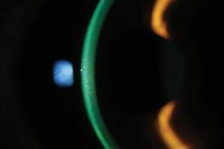
| Epi-on CXL with riboflavin completely penetrating to posterior cornea. Photo: Andrew Morganstern, OD |
The procedure has been less successful in patients with previous RK surgery. As the wounds are frequently unstable, crosslinking does not have the same impact that it typically produces in a normal keratoconic patient. Some centers outside of the United States will treat both the keratoconus with CXL and the current refractive error using topo-guided wavefront laser systems and PRK treatments to simultaneously allow for both visual rehabilitation and stabilization.
CXL in the Clinic
The procedure is relatively straightforward: if the patient is epi-on, the riboflavin is administered every two minutes until there is full corneal saturation and riboflavin “flare” in the anterior chamber is achieved upon physical slit-lamp assessment by the clinician. In most patients, this is typically achieved in 30 to 40 minutes; however, it may take longer in younger individuals because the corneal epithelial matrix is more difficult for the riboflavin to penetrate. The epi-off method uses the same procedure, but 30 minutes is typically sufficient in most cases to provide full saturation before initiation of treatment. Most riboflavin CXL applications are done one eye at a time, but some newer technologies offer simultaneous bilateral treatment.
The postoperative course of the patient is predicated on which of the two interventions is selected for the individual. Epi-off patients are typically placed in a bandage contact lens (BCL), and topical antibiotics and steroids are used QID for one week—much like post-PRK management. Once epithelial closure has been achieved, the antibiotic is stopped and the steroid is tapered over three additional weeks. In my experience, the wound healing is slightly slower than we typically see in a PRK treatment, and may last up to seven days or longer post-intervention.
Epi-on patients also receive a BCL plus topical antibiotics and steroids; the anti-inflammatory is then tapered over several weeks. The antibiotic is typically stopped in three days, once the epithelium has completely healed.
During epi-on treatment, patients usually experience some discomfort in the first 24 hours due to the toxicity from the numerous drops applied during the procedure and the UV light. This discomfort usually dissipates by the next morning. Additionally, to blunt the discomfort, I typically use homatropine 5% preoperatively and postoperatively along with a topical NSAID. In my experience with epi-on, the BCL can be removed the following day, and the patient can return to normal activities at that time.
In most cases, because the vision is not optimal with correction prior to the procedure, acuity is only marginally affected and not a noticeable part of the postoperative course. Epi-on patients will have minimal, if any, complications beyond the noticeable corneal haze in some patients.
Post-op Complications and Lens Wear
Predominantly observed in the epi-off treatment protocol, these can involve delays in wound healing, microbial keratitis and stromal haze development.
I have observed several epi-off patients who present with wound healing delays and increased discomfort. Oftentimes, these complications resolve within seven days, but it is not unusual—even with continued use of a BCL—that a defect may remain for a more prolonged period. In cases such as this, I have had excellent success with pressure patching and the use of a steroid antibiotic unguent (e.g., Tobradex) for one or two days.
Most epi-on patients can return to contact lens wear within one week, if wearing soft lenses or combination, piggyback fits, but we would recommend 10 to 14 days for hybrid lenses and more complex interventions. In patients who have undergone the epi-off procedure and plan to resume lens wear, for soft lenses my recommendation is seven to 14 days; for hard lens wear, approximately three to four weeks; and for complex lens wear, a four- to six-week period. This is due in part to the experiences that I have had with the epithelial remodeling after the procedure, and the potential for a recurrent erosion-like presentation if the cornea is manipulated significantly in the early postoperative course.
It is important to remember that the crosslinking process can continue for up to six months in most patients, which can produce a relative flattening and thinning of the cornea that may affect lens design. Typically, this will be accompanied by a light haze that indicates active crosslinking.
Corneal collagen crosslinking will (literally) alter the landscape of keratoconus once it’s approved in the US. Its potential to produce a cessation of disease progression, and to improve the visual welfare of the treated individual, is unrivaled.
Because the procedure is best for patients who show true progression of their keratoconus, it should be limited to these individuals. As such, the minimum age for the procedure is 10; the maximum age typically tails out at 40 to 45 years, as the body crosslinks naturally secondary to ultraviolet light exposure and the body’s own blood sugar levels. This accounts for the often-noted cessation of progression in a keratoconic patient as they enter their fourth to fifth decade of life. While this is true of the vast majority of cases, I have seen several individuals older than 45 with progressive disease—typically resulting from trauma.
CXL typically provides a change in the refractive error in a significant percentage of patients. Most will show a mild decrease in myopia, astigmatism or both over the first three to six months while the crosslinking process is completed. As the science surrounding the treatment has grown both internationally and in US clinical trials, corneal collagen crosslinking has the potential to forever alter the impact of keratoconus globally.


