 |
A 17-year-old female presented for a contact lens and ocular health exam. She said she had noticed a “bubble” on her eye. It was not painful but she stated she has been “trying to pop it.” She reported no change in her vision and her slit lamp exam was otherwise unremarkable. She was diagnosed with conjunctival lymphangiectasis and was told to stop trying to remove it, as it is a normal part of her eye.
Conjunctival lymphangiectasis is a condition wherein subconjunctival fluid accumulates from dilated conjunctival lymphatic channels. It can be seen most readily on the bulbar conjunctiva.1 The bubble will often have a cystic appearance and may be clear or yellow. Elevated lymphatic channels are separated by translucent septate walls.
The etiology is unknown, but the condition is presumed to involve obstructed lymphatic channels.
There is one report in the literature that defines conjunctival lymphangiectasis as an under-recognized ocular sign in the presence of Fabry’s disease.1 Although conjunctival lymphangiectasis is considered benign, clinicians should remain vigilant for signs of bilateral corneal verticillata in order to rule out Fabry’s.
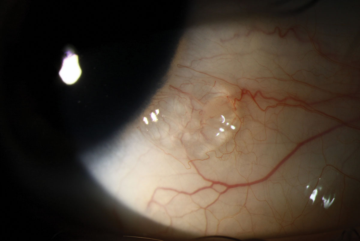 |
| Conjunctival lymphangiectasis can present as a bubble on the bulbar conjunctiva that often has a cystic appearance and may be clear or yellow. Click image to enlarge. |
| 1. Sivley MD, Wallace EL, Warnock DG, Benjamin WJ. Conjunctival lymphangiectasia associated with classic Fabry disease. Br J Ophthalmol. 2018 Jan;102(1):54-58. |

