 |
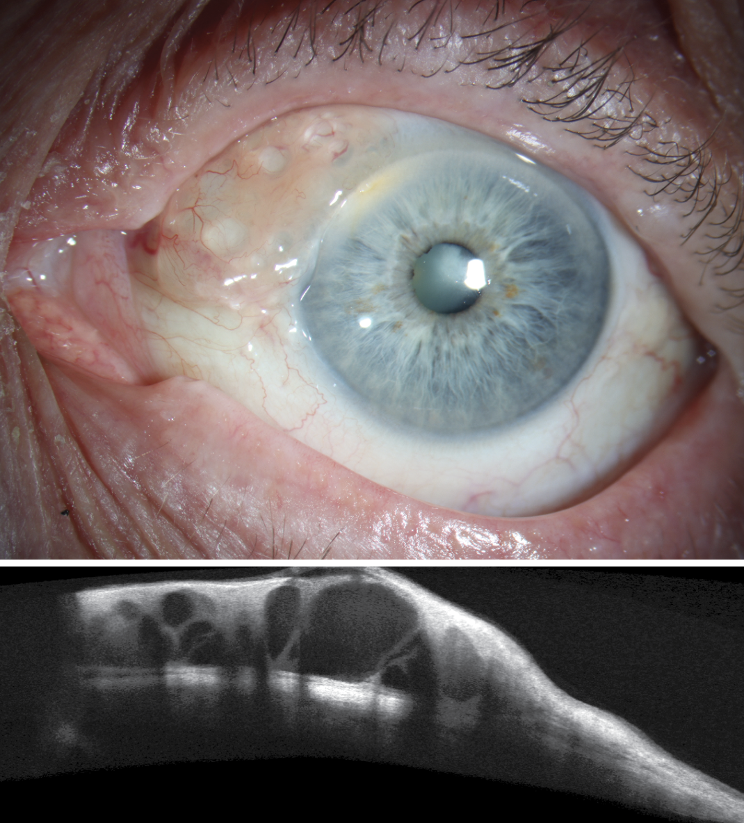
An 84-year-old man presented for evaluation of conjunctival lesion OS with concerns for potential malignancy. The patient reported the lesion “had been there for years,” and he denied discomfort. The patient was diagnosed with a large multi-lobular conjunctival cyst and monitored yearly.
Conjunctival cysts are benign fluid-filled sacs. They vary in size and may be asymptomatic or cause symptoms such as discomfort, irritation or foreign body sensation. The pathogenesis is multifactorial, involving mechanical, developmental and inflammatory processes that disrupt the normal cellular architecture of the conjunctiva.
One of the most common mechanisms is blockage of the superficial or deep lymphatic drainage system, leading to fluid accumulation within the cyst, which is typically thin and clear. The pressure within the cyst can eventually cause it to become more prominent, leading to noticeable swelling or bulging on the conjunctiva.
Another important pathway involves the entrapment and proliferation of epithelial cells. A cyst forms as squamous epithelial cells lining the conjunctiva undergo excessive proliferation and eventually form a sac-like structure. Over time, these cells secrete fluid, causing the cyst to enlarge. The cystic structure filled is with cellular debris, tears and sometimes inflammatory exudates. This may be congenital but often happen after trauma or surgical procedures, such as cataract surgery.
 |
|
Click image to enlarge. |
Inflammation and infection can also contribute to development of conjunctival cysts. Bacterial or viral conjunctivitis, blepharitis or chronic dry eye disease can lead to localized inflammatory responses that may impair normal glandular function or damage the conjunctival epithelium and lead to the formation of cysts.
In some cases, conjunctival cysts can be congenital. These typically form during fetal development, when epithelial remnants from the embryonic stage fail to undergo proper regression or differentiation. As a result, epithelial-lined cysts are left behind within the conjunctiva.
Other potential contributing factors include age-related changes in the conjunctiva, mechanical trauma and autoimmune disorders. These conditions can alter the normal drainage and function of the conjunctival structures, leading to cyst formation.
Treatment, when needed, often involves aspiration or surgical removal, usually with a low risk of recurrence.
It is always wise to differentiate a cyst from a more malicious lesion such as conjunctival neoplasm. While cysts are smooth, round and fluid-filled, neoplasms are solid and irregular with variable pigmentation.
OCT imaging of our patient showed superficial fluid-filled vacuoles, which remained stable over time. Since he was comfortable, no treatment was indicated.


